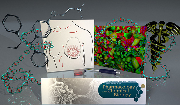
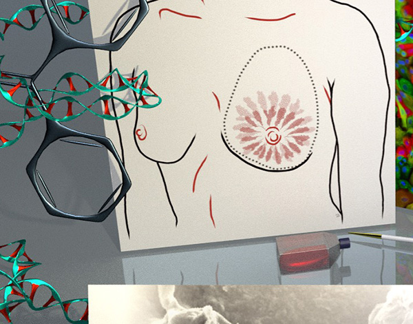
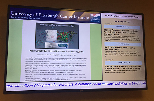
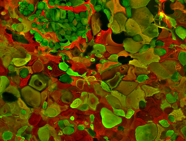
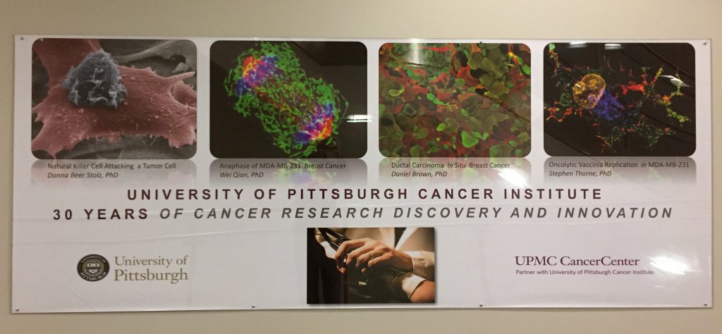
This is a little graphic I made for an RFA (request for application) for grant submissions (with breast cancer-related imagery). This work also contains within it my previous works from a series I took for “Images Fighting Cancer” and a Breast Diagram.
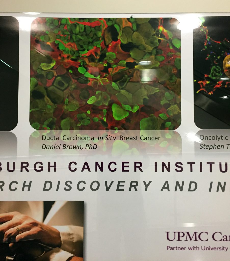
The image also showed up on the University of Pittsburgh Cancer Institute (UPCI) television screens around campus.
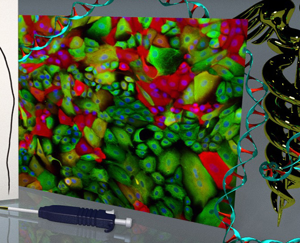
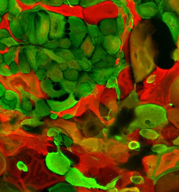
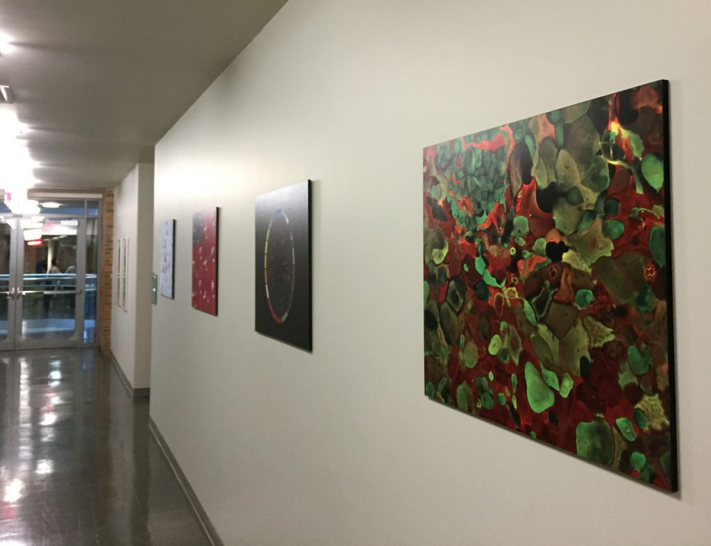
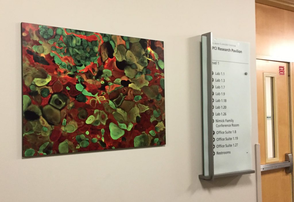
I take a lot of images of cells fluorescently stained via immunocytochemistry. I submitted this one for an “Images fighting cancer” competition, with the winners being chosen to be printed and displayed at Hilman Cancer Center. Mine was one of the images chosen!
These are cells derived from human DCIS (early stage breast cancer) stained for cytokeratins 8 and 14.
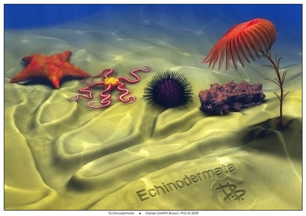
From August 2009 to August 2010 I worked as a post-doc in the lab of Dr. Veronica Hinman at Carnegie Mellon University. Basically, I studied the evolution of gene regulatory networks (GRNs). Specifically, our lab focused on looking at GRNs in the context of development using the wonderful sea critters in the phylum Echinodermata. For those of you not in the know, the “spiny-skinned” echinoderms are the asteroids (starfish/sea stars), ophiuroids (brittle stars), echinoids (sea urchins), holothuroids (sea cucumbers), and crinoids (feather stars, sea lillies and such).
In celebration, I spent a fair bit of time getting back to my art roots creating the above cladogram in the sand of the Echinoderm phylum.
I spent a while trying to find time-lapses or animations of starfish development online, to no avail. Thus I spent a week of much needed downtime to create this computer animation using Blender: (note – you can also watch it in High Definition on youtube).
NOTE: The details of the actual metamorphosis of the rudiment into the juvenile are not accurate – it’s quite hard to animate these types of changes – and to be honest I had not actually seen these creatures in the flesh. But it’s good enough to get a good idea of how the whole developmental process occurs in this type of sea star.
Way back in 2005/2006 I was trying to finish up a Ph.D. in biology studying the genetics of heart development in the lab of Dr. Frank Conlon at UNC, using the frog as a model. My advisor at the time was also going up for tenure and wanted a decent animation of heart development for his presentation. Thus I convinced him to buy us a copy of Maya, with which I made the following video and started learning how to create 3D art and animations.
The Cretacious-Tertiary Boundary
Sixty-five million years ago, a daily struggle occurs in the midst of the world-changing event that would result in the demise of the non-avian dinosaurs and the rise of our own lineage of mammals. The layer of rock demarkating the end of the Cretaceous and beginning of the Tertiary is known by geologists as the “K-T Boundary.” The mammals in this case are “cynodonts” – our ancestors in the late Cretaceous. This took me three weeks to create, using Blender and GIMP software packages.
“K-T” Detail
CLICK HERE to see how this piece (and generally all my 3D art) is made.